
Image Production · Exposure and contrast2 min read5-question check
Exposure Latitude in Digital Radiography
DR forgives exposure errors, but a good-looking image can still mean too much dose.
The rule
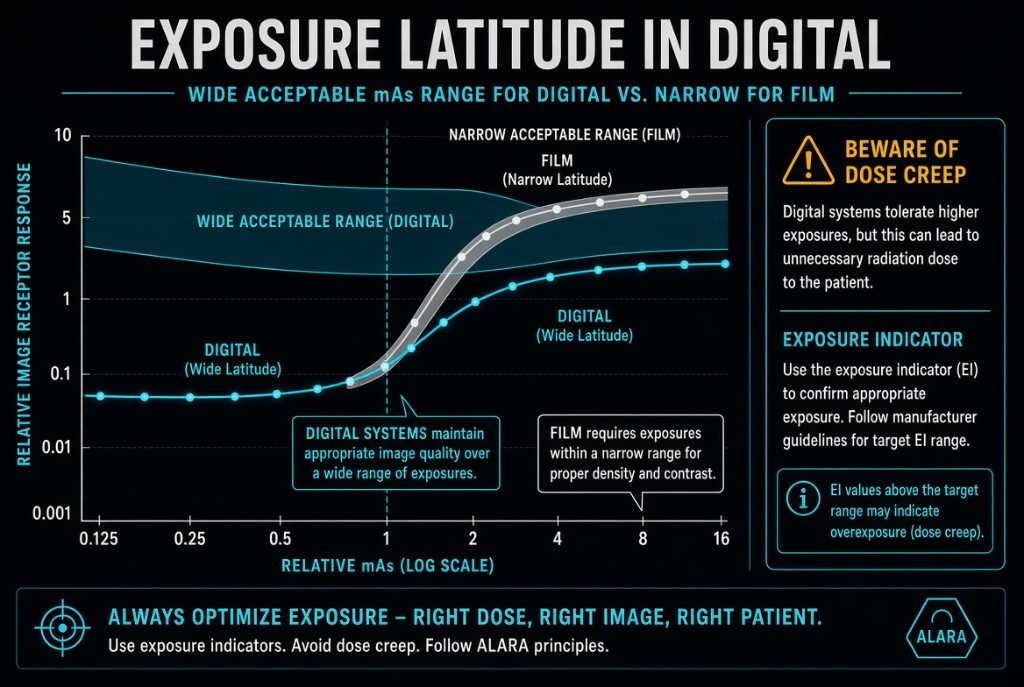
Exposure latitude is the range of exposures that still produce a diagnostic image. Digital detectors have a wide exposure latitude. Processing rescales the signal, so an image made with too much or too little exposure can still appear with normal brightness. Film-screen systems had a narrow latitude, and errors showed up immediately as a film that was too light or too dark.
Exposure Latitude
How it works
A digital detector responds in a linear way over a very large range of exposure. The computer finds the useful signal in the histogram and maps it to standard display values. That rescaling is why brightness on the monitor tells you almost nothing about how much radiation the detector received.
The limits
The latitude has edges. Underexposure leaves too few photons, and the image shows quantum mottle, a grainy noise that processing cannot remove. Overexposure produces a clean, low-noise image, so it rarely looks wrong. Only with large overexposure does the detector saturate, and the areas receiving the most exposure, such as thin anatomy and skin edges, lose all detail and display as uniform black. Between those edges, the image looks fine while the patient's dose may be far higher than needed.
On the job
Wide latitude is a safety net for small technique errors, especially on mobile exams. It is not permission to overexpose. If a radiographer routinely doubles mAs to avoid mottle, the images still look good, but every patient receives twice the dose. This gradual upward drift is called dose creep. The exposure indicator is the routine check on receptor exposure, so review it on every image.
Clinical Example
A student notices that a room's chest technique has crept up to twice the protocol mAs over several months. The images look excellent, so nobody complained. The exposure indicators have been running high, and returning to the protocol technique produces equally diagnostic images at half the dose.
Common Mistakes
- Judging exposure by image brightness, which processing has already corrected.
- Treating wide latitude as permission to add mAs to avoid any chance of noise.
- Expecting processing to fix an underexposed image; it cannot remove quantum mottle.
- Expecting a moderately overexposed digital image to look dark, the way film did.
Why It Matters
Wide latitude lowers repeat rates, but it hides overexposure and lets dose creep go unnoticed. The registry tests what latitude means, why dose creep happens, and what underexposure and overexposure look like in DR.
Key Relationships
- Wide latitude → fewer repeats, but overexposure is hidden
- Underexposure → quantum mottle that processing cannot fix
- Double the mAs → double the dose, with little visible change
- Large overexposure → detector saturation and loss of detail