
Procedures · Extremities3 min read5-question check
Elbow and forearm film check
Open joint on the AP, a lateral flexed 90° with concentric circles, radial head free on the lateral oblique, coronoid in profile on the medial oblique.
The AP
On a good AP elbow, the arm is fully extended and the hand supinated, with the epicondyles parallel to the IR. The elbow joint is open and the radial head overlaps the proximal ulna just a little. If the patient cannot extend the elbow, take two APs instead of forcing it: one with the humerus flat on the IR and one with the forearm flat.
The lateral
The lateral elbow is flexed 90 degrees with the hand in a true lateral, thumb up. Lower the shoulder so the humerus and forearm lie on the same level as the IR. On a good lateral, three concentric arcs show at the distal humerus: the trochlear sulcus, the capitulum and the medial ridge of the trochlea. The olecranon is in profile and the joint is open. Flexion matters for more than looks: at 90 degrees the posterior fat pad is normally hidden, so a visible posterior fat pad suggests a fracture. An elbow left partly extended can show that fat pad when there is no fracture.
The two obliques
The AP lateral oblique, also called the external oblique, turns the hand and arm out about 45 degrees. It frees the radial head, neck and tuberosity from the ulna. The AP medial oblique, or internal oblique, turns the hand in, palm down. It shows the coronoid process in profile, with the radial head now behind the ulna. If you cannot remember which is which, look at the radial head: free of the ulna means lateral oblique.
The forearm
The AP forearm includes both the wrist and elbow joints, with the hand supinated and the epicondyles parallel to the IR. If the hand is pronated, the radius crosses over the ulna, which hides the proximal radius and the radial head. On the lateral forearm, the elbow is flexed 90 degrees and the distal radius and ulna superimpose, just like the lateral wrist.
Read the films
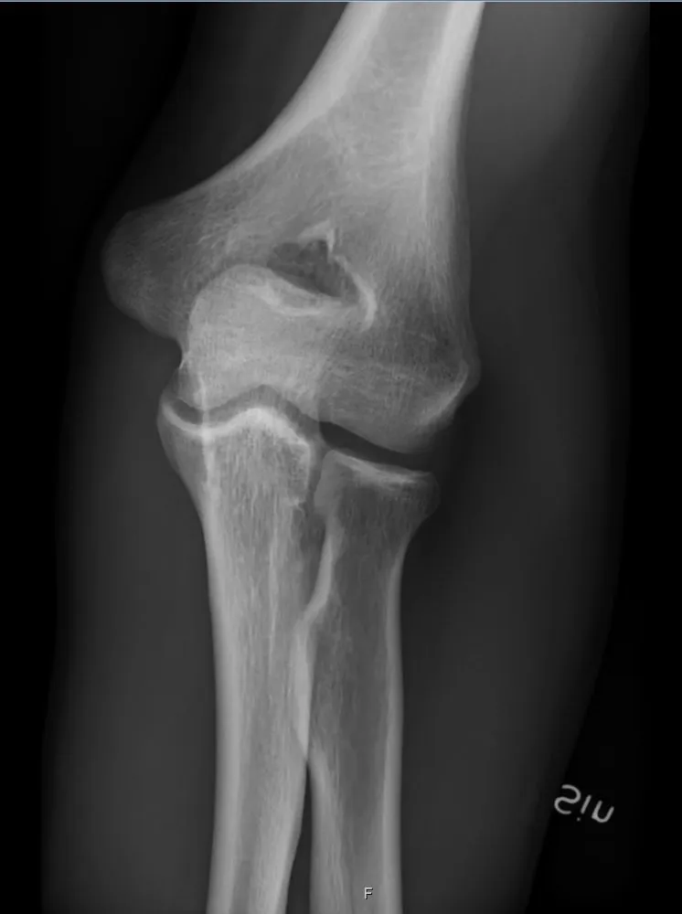
KeeperAP elbow: a keeper
Look for: The joint is open, the epicondyles are seen on each side, and the radial head overlaps the proximal ulna just a little.
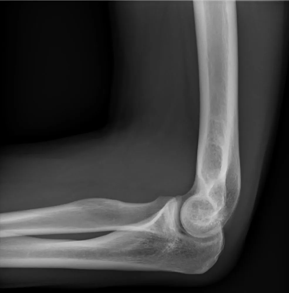
KeeperLateral elbow: a keeper
Look for: Flexed 90 degrees, concentric arcs at the distal humerus, the olecranon in profile and an open joint.
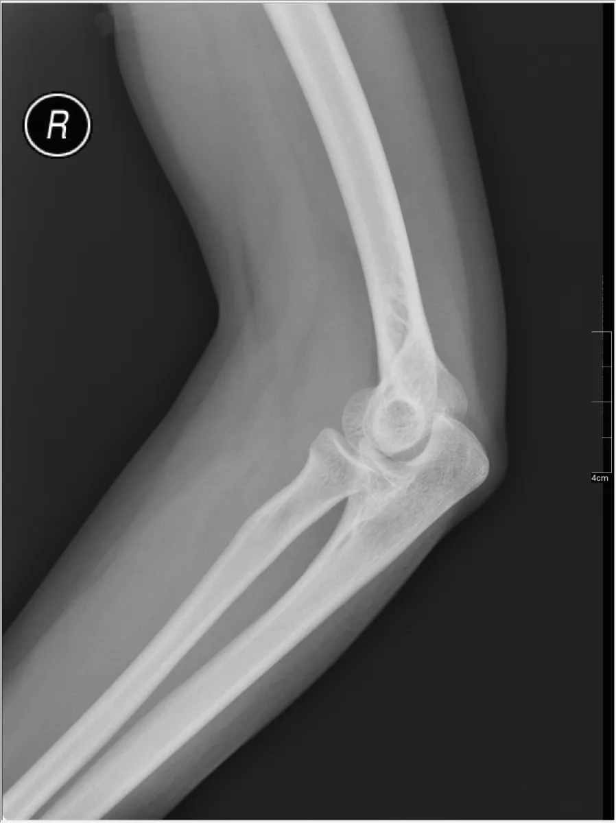
RepeatLateral elbow, not flexed
Look for: The elbow is bent only partway. The fat pads cannot be judged properly and the joint is not shown the standard way.
Fix: Flex the elbow 90 degrees with the hand in a true lateral, and lower the shoulder so the humerus and forearm are on the same level.
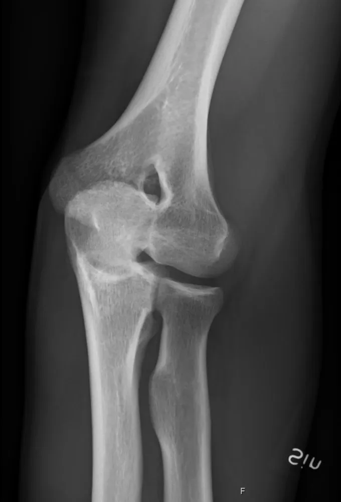
KeeperAP lateral (external) oblique: a keeper
Look for: The radial head and neck are free of the ulna. That is how you know it is the lateral oblique.
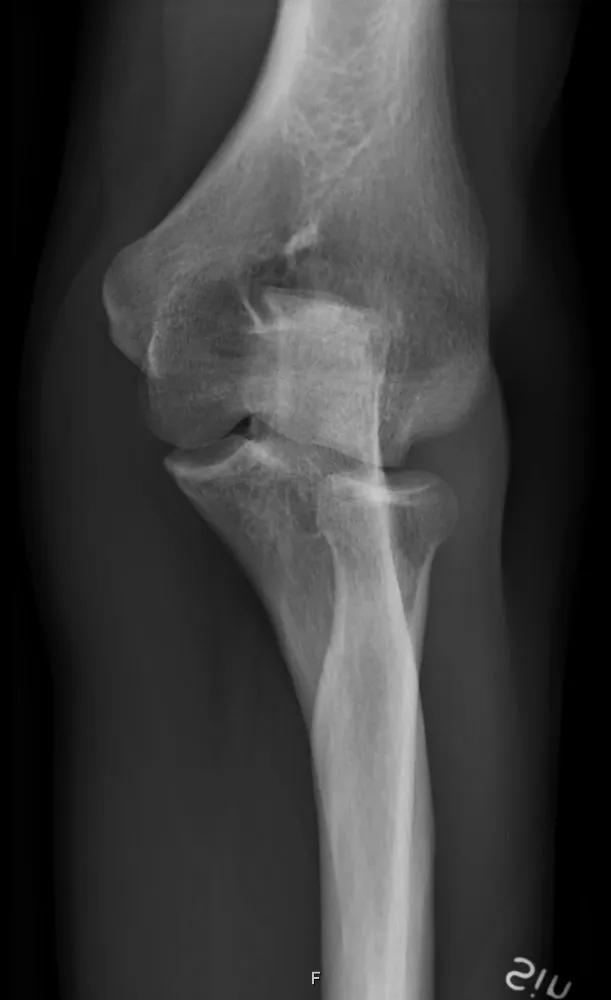
KeeperAP medial (internal) oblique: a keeper
Look for: The coronoid process is in profile and the radial head sits behind the ulna.
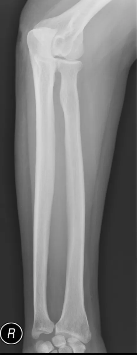
RepeatAP forearm, radius crossed over the ulna
Look for: Near the elbow the radius crosses over the ulna, so the radial head and neck are hidden. The hand was turned toward pronation.
Fix: Supinate the hand fully and set the epicondyles parallel to the IR.
Clinical Example
A child with a fall on an outstretched hand cannot straighten the elbow. You skip forcing a straight AP. You take an AP with the humerus flat and another with the forearm flat, then a lateral at 90 degrees. The lateral shows a raised anterior fat pad and a visible posterior fat pad, which you point out to the radiologist.
Common Mistakes
- Leaving the shoulder high on the lateral, so the humerus is not on the same level as the forearm.
- Taking the lateral only partly flexed, which can show a posterior fat pad when there is no fracture.
- Mixing up the obliques; the lateral oblique frees the radial head.
- Pronating the hand on the AP forearm, which crosses the radius over the ulna.
Why It Matters
Elbow fractures in children are often seen only as a fat pad sign, and that sign only works on a true lateral flexed 90 degrees.
Key Relationships
- Lateral: 90° flexion, concentric arcs at the distal humerus
- Posterior fat pad visible at 90° → suspect fracture
- Lateral (external) oblique → radial head free
- Medial (internal) oblique → coronoid in profile
- Pronated forearm → radius crosses the ulna