
Procedures · Extremities2 min read5-question check
Elbow and forearm
AP supinated, lateral at 90 degrees with concentric rings, and obliques for the coronoid and radial head.
Patient and part
Lower the shoulder so it lies in the same plane as the elbow. For the AP elbow, extend the elbow fully, supinate the hand and keep the humeral epicondyles parallel to the IR. For the lateral elbow, flex to 90 degrees, place the epicondyles perpendicular to the IR and turn the hand lateral, thumb up. For the AP oblique with medial rotation, pronate the hand and rotate the arm about 45 degrees medially. For the AP oblique with lateral rotation, rotate the arm about 45 degrees laterally. The forearm is always taken AP with the hand supinated, never PA, because pronation crosses the radius over the ulna. The AP and lateral forearm must include both the wrist and elbow joints.
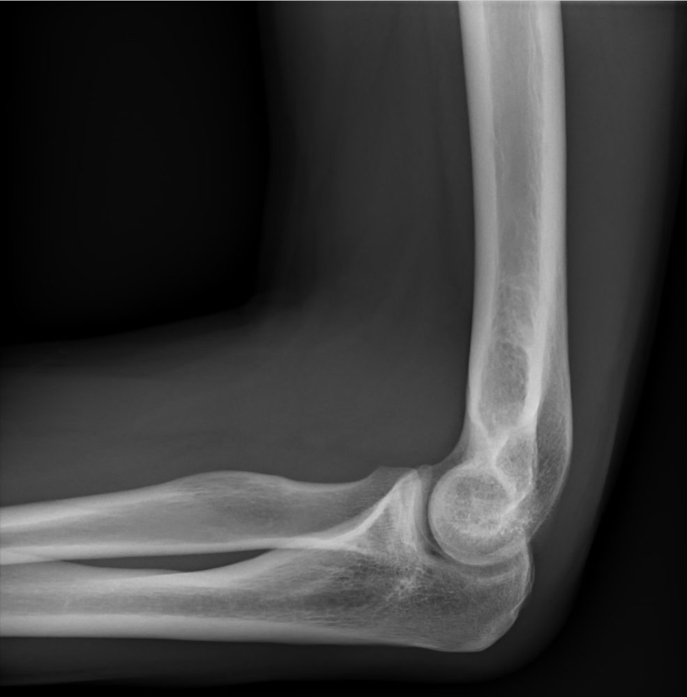
Lateral elbow at 90 degrees
Central ray
Use 40 inches, about 100 cm. For every elbow view, the CR is perpendicular to the mid elbow joint. For the forearm, the CR is perpendicular to the midforearm, with the IR long enough to include both joints.
What a good image shows
On the AP elbow, the joint is open and the radial head, neck and tuberosity slightly overlap the proximal ulna. On the lateral, three concentric arcs appear at the distal humerus, the olecranon is in profile, the joint is open and the radial head partly overlaps the coronoid process. The medial rotation oblique shows the coronoid process in profile. The lateral rotation oblique shows the radial head, neck and tuberosity free of the ulna. On a true lateral, the posterior fat pad is normally hidden in the olecranon fossa. A visible posterior fat pad, or an anterior fat pad lifted like a sail, signals joint effusion and suggests a hidden fracture.
Fixing it
If the arcs on the lateral are not concentric, the epicondyles were not superimposed. A common cause is a shoulder higher than the elbow, so lower it into the same plane. If the elbow cannot fully extend, take two APs instead of one, one with the humerus parallel to the IR and one with the forearm parallel.
Clinical Example
A child fell on an outstretched arm and the AP elbow looks normal. On the true lateral, the posterior fat pad is visible, and the radiologist reads an effusion from a likely occult supracondylar fracture.
Common Mistakes
- Taking the forearm PA, which crosses the radius over the ulna.
- Raising the shoulder above the elbow, which breaks up the concentric arcs.
- Mixing up the obliques: medial rotation shows the coronoid, lateral rotation shows the radial head.
- Including only one joint on a forearm study.
Why It Matters
A true lateral is the only view that reliably shows fat pad signs, which often reveal a fracture you cannot see directly. The registry tests which oblique shows which structure and what concentric rings mean.
Key Relationships
- Lateral elbow: 90 degrees flexion, epicondyles perpendicular to IR
- Medial rotation oblique: coronoid process in profile
- Lateral rotation oblique: radial head free of ulna
- Visible posterior fat pad: effusion, suspect fracture
- Forearm: AP, supinated, both joints