
Safety · Radiobiology3 min read5-question check
Dose-response and cell survival curves
Linear no-threshold guides protection, the S-shaped threshold curve fits tissue reactions, and the shoulder on a cell survival curve is repair.
Four curve shapes
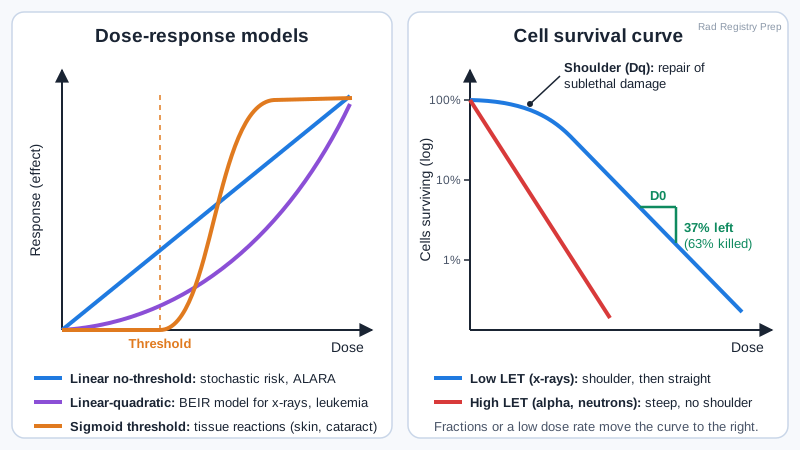
A dose-response curve plots a biologic effect against dose, and the registry expects you to match each shape to its effects. Linear no-threshold (LNT): a straight line from zero, so any dose carries some risk and doubling the dose doubles the risk. LNT is the conservative model behind radiation protection standards and ALARA. Linear-quadratic no-threshold: the line curves upward, rising slowly at low doses and faster at high doses. The BEIR committee uses it for low-LET radiation such as x-rays, especially for leukemia. Linear threshold: no response until a set dose, then a straight-line rise; it is the least used. Nonlinear threshold, also called sigmoid or S-shaped: nothing below the threshold, a steep rise, then a plateau where nearly everyone is affected. It fits deterministic effects (tissue reactions) such as skin erythema, epilation, cataracts and blood count depression.
Dose-response models and the cell survival curve
Cell survival
A cell survival curve plots the fraction of cells still able to divide against dose, with survival on a logarithmic scale. For x-rays, which are low-LET, the curve starts with a shoulder: at low doses cells repair sublethal damage, so few die. The width of the shoulder is the quasi-threshold dose (Dq). After the shoulder the line is straight, and each additional mean lethal dose (D0) leaves only 37% of the remaining cells alive, killing 63%. Mammalian cells have a D0 of roughly 1 to 2 Gy. High-LET radiation such as alpha particles and neutrons gives a steeper line with almost no shoulder, because its dense ionization damages DNA beyond repair.
Dose rate and fractions
The same total dose does less harm when it is spread out, because cells repair between doses. Fractionation divides the dose into equal parts with time in between, as in radiation therapy, where normal tissue recovers between treatments. Protraction delivers the dose continuously at a low dose rate. Both cause less damage than one large dose given quickly. Oxygen makes cells more sensitive: well-oxygenated cells need less dose for the same kill, and the oxygen enhancement ratio for x-rays is about 3. Hypoxic tumor cells are the most resistant.
On the job
Diagnostic doses sit at the very bottom of the LNT line, far below any threshold. You will never see a tissue reaction from a routine radiograph, but the LNT model says the small stochastic risk is never zero. That is why you collimate, avoid repeats and keep your distance on every exam, and why long fluoroscopy cases, which can cross skin thresholds, are tracked with air kerma and DAP.
Clinical Example
After a 90-minute interventional case, a patient develops reddened skin on the back two weeks later. The skin dose passed the threshold on a sigmoid, deterministic curve. The same patient's small added cancer risk is plotted on the LNT line and cannot be predicted for any one person.
Common Mistakes
- Matching cancer and leukemia to a threshold curve; stochastic effects use no-threshold models.
- Thinking the shoulder on a cell survival curve means radiation does nothing at low dose; it shows repair of sublethal damage.
- Assuming one large dose and the same dose in fractions do the same damage; fractionation and protraction reduce damage.
- Saying high-LET radiation has a broad shoulder; it has little or none because its damage is not repaired.
Why It Matters
Curve shapes explain why diagnostic exposures never cause tissue reactions yet still require ALARA, and why radiation therapy is given in fractions. The registry asks you to match effects to curves and to read a cell survival curve.
Key Relationships
- LNT → basis for radiation protection, stochastic risk
- Linear-quadratic → BEIR model for low-LET, leukemia
- Sigmoid threshold → deterministic effects (skin, cataract, blood)
- Shoulder (Dq) → sublethal damage repair; none for high LET
- D0 → leaves 37% alive (kills 63%)
- Fractionation and protraction → less damage for the same total dose
- OER for x-rays ≈ 3; oxygen increases radiosensitivity