
Image Production · Digital imaging and processing2 min read5-question check
Digital windowing
Windowing changes how the image is displayed, not the data or the dose, and cannot rescue a bad exposure.
The rule
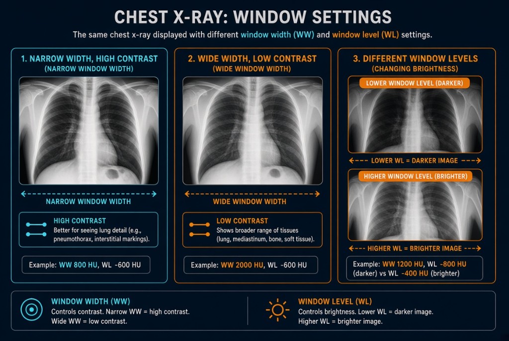
Windowing is post-processing at the display. After the system has processed the acquired image, the radiographer or radiologist can change how its pixel values are shown as shades of gray on the monitor. Window level adjusts overall brightness. Window width adjusts contrast. Neither changes the exposure the patient received, and neither changes the image data that was acquired.
Window Width and Window Level
How it works
Each pixel holds a number. A monitor can show only a limited range of gray shades, so the window selects which range of pixel values is spread from black to white. Values outside the window are clipped to pure black or pure white. Move the window, and a different slice of the data gets the full gray scale. Narrowing it raises contrast; widening it lowers contrast and shows more grays. The exact effect of each control is a separate lesson on window width and level.
The limit
Windowing redistributes what is already in the data. It cannot add photons. An underexposed image has quantum mottle built in, and adjusting brightness or contrast often makes the noise more obvious, not less. An area overexposed enough to saturate the detector has no detail left to bring back. Windowing also cannot fix motion, poor positioning, or anatomy that was clipped by collimation.
On the job
Because automatic rescaling and windowing can make almost any exposure look acceptable, overexposure is easy to miss. Over time, this leads to dose creep, where techniques drift upward because no image ever looks too dark. Judge exposure by the exposure indicator, not by how the image looks after adjustment. Use windowing to help show the anatomy, and follow department policy before saving an adjusted image to PACS.
Clinical Example
A lateral lumbar spine looks grainy, so a student widens the window and lowers the brightness until it looks smoother. The deviation index still reads minus 3. The image is underexposed, and windowing cannot add the missing photons, so the student repeats with a corrected technique.
Common Mistakes
- Thinking windowing changes patient dose or the acquired data.
- Trying to fix an underexposed, noisy image by windowing.
- Judging exposure by appearance after windowing instead of by the exposure indicator.
- Mixing up the controls: level is brightness, width is contrast.
Why It Matters
Windowing makes images easier to read, but it can also conceal exposure errors that cost the patient dose. The registry tests what windowing can and cannot change.
Key Relationships
- Window level → brightness
- Window width → contrast
- Windowing → no change in dose or acquired data
- Windowing cannot add photons or recover saturated detail