
Image Production · Exposure and contrast2 min read5-question check
Receptor exposure vs contrast
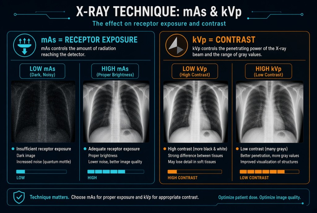
mAs sets how many photons reach the receptor; kVp sets penetration and subject contrast.
The rule
Receptor exposure is the amount of radiation striking the image receptor. Film texts called its visible result density. In digital radiography, mAs is the primary controller of receptor exposure: double the mAs and twice as many photons reach the detector. But the computer rescales brightness during processing, so a wrong mAs does not show up as a dark or light image. Too little shows up as noise. Too much shows up as unneeded patient dose and, at the extreme, saturation. The exposure indicator is how you see it.
Receptor Exposure and Contrast
Why it happens
kVp controls beam energy and penetration, which sets subject contrast, the difference in radiation leaving different tissues. Low kVp exaggerates the absorption difference between bone and soft tissue, giving high contrast with few grays, a short scale. High kVp penetrates tissues more evenly, giving lower contrast with many grays, a long scale, and a larger share of scatter. In digital imaging, displayed contrast is also shaped by processing and windowing, but kVp still decides how much tissue difference exists in the data.
The numbers
kVp also changes receptor exposure, and strongly. By the 15 percent rule, raising kVp 15 percent roughly doubles receptor exposure, and lowering it 15 percent roughly halves it. Worked example: a knee at 70 kVp and 10 mAs. Raise kVp 15 percent to about 80 and cut mAs to 5. Receptor exposure stays about the same, contrast is a little lower, and patient dose drops.
On the job
Choose kVp first: high enough to penetrate the part, at the contrast the exam needs. A chest uses high kVp so lungs, heart, and ribs all show; extremities use lower kVp for bony detail. Then choose mAs to give the receptor the right number of photons. Afterward, judge mAs by the exposure indicator, not by how bright the image looks.
Clinical Example
A lateral knee comes up with a good-looking image, but the deviation index is plus 3. The receptor received twice the target exposure. Next time you keep 70 kVp and halve the mAs, or raise kVp 15 percent and cut the mAs to one quarter.
Common Mistakes
- Expecting a digital image to look dark when mAs is too high.
- Saying kVp has little effect on receptor exposure, when a 15 percent change doubles or halves it.
- Confusing high kVp with high contrast, when higher kVp lowers contrast.
- Raising mAs to fix contrast instead of adjusting kVp.
Why It Matters
Choosing kVp and mAs correctly gives a diagnostic image at the lowest dose. The registry tests which factor is primary for each and the 15 percent rule math.
Key Relationships
- Double mAs → double receptor exposure
- Raise kVp 15 percent → about double receptor exposure
- Higher kVp → lower contrast, more grays
- Too little exposure → noise; too much → dose and saturation