
Image Production · Digital imaging and processing2 min read5-question check
Direct vs indirect DR detectors
Indirect DR turns x-rays into light, then charge. Direct DR turns x-rays straight into charge with selenium.
Two kinds
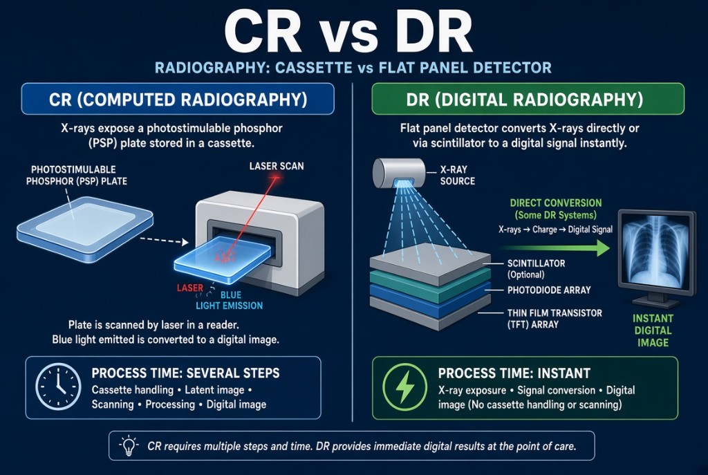
Digital radiography flat-panel detectors come in two kinds, indirect and direct. Both produce an image within seconds without a separate reader, and both read out through a grid of detector elements called DELs, each with its own thin-film transistor, or TFT, that acts as a switch. The difference is how the x-ray energy becomes an electric signal. Computed radiography is no longer on the 2027 registry exam.
CR vs DR
Indirect conversion
An indirect detector uses two steps. First, a scintillator converts x-rays into light. The common scintillators are cesium iodide and gadolinium oxysulfide. Second, an amorphous silicon photodiode in each DEL converts that light into electric charge, which the TFT sends out to be digitized. Some indirect systems use a CCD or CMOS sensor in place of the photodiode array. Because light spreads a little in the scintillator before reaching the photodiode, sharpness is slightly reduced. Cesium iodide is grown in needle-shaped crystals that channel the light and limit this spread.
Direct conversion
A direct detector uses one step. A layer of amorphous selenium, a photoconductor, absorbs the x-rays and produces electric charge directly. An electric field across the selenium pulls the charge straight down to the collecting electrode, so there is very little sideways spread. Amorphous selenium direct detectors are widely used in digital mammography, where fine detail is critical.
In practice
In both types, spatial resolution is limited by DEL size: smaller DELs give finer detail. Fill factor is the share of each DEL that is actually sensitive to the signal; the rest holds the TFT and wiring. Smaller DELs usually have a lower fill factor, so they may need more exposure for the same signal. Both detector types need correct receptor exposure, report an exposure indicator, and show quantum mottle when underexposed. Wireless panels are fragile and must be handled with care.
Clinical Example
A department replaces an older gadolinium oxysulfide wall bucky panel with a cesium iodide indirect detector. After testing, the physicist lowers the technique chart slightly. She explains that cesium iodide absorbs x-rays efficiently and its needle structure limits light spread.
Common Mistakes
- Calling amorphous silicon the x-ray absorber in an indirect detector; it detects light from the scintillator.
- Thinking direct detectors need a scintillator.
- Assuming DR detectors do not need correct technique because the image is processed.
- Mixing up the TFT, which is a switch, with the photodiode, which converts light.
Why It Matters
Knowing the conversion steps explains differences in sharpness, dose efficiency, and artifacts. The registry asks which materials belong to which detector type.
Key Relationships
- Indirect: x-ray → light (scintillator) → charge (a-Si photodiode) → TFT
- Direct: x-ray → charge (amorphous selenium) → TFT
- Smaller DEL → better spatial resolution
- Light spread in the scintillator → slight loss of sharpness