
Procedures · Chest, abdomen and GI4 min read5-question check
Chest film check
Before the patient leaves, check the film: whole lungs, no rotation, 10 ribs, clear apices, and on the lateral, ribs that line up.
The 10-second check
Every chest gets the same quick check before the patient leaves the room. Are the whole lungs on the film, from the apices to both costophrenic angles and out to both lateral rib margins? Is the patient straight? Did they take a full breath? Are the scapulae out of the lungs and the clavicles below the apices? Is anything lying over the apices, like the chin or the arms? Is the exposure indicator in range, with the spine faintly visible through the heart? The real films below show a good PA and lateral next to the misses you will see most.
Rotation and inspiration
Rotation shows at the top of the film. Find the medial ends of both clavicles and the spinous process between them. On a straight PA the two clavicle ends sit the same distance from the spine. If one is farther away, the patient was turned, the heart and trachea shift, and the heart size cannot be judged. Square both shoulders against the IR and check that weight is even on both feet. Inspiration shows at the bottom. Count the posterior ribs above the right hemidiaphragm. Ten is the goal. Fewer means a shallow breath, which crowds the lung markings and can mimic disease. Expose at the end of the second deep breath, not the first.
Field, chin and shoulders
The field runs from about 1.5 to 2 inches above the shoulders to below the costophrenic angles, with the CR at T7, about 7 to 8 inches below the vertebra prominens. Small effusions collect first in the costophrenic angles, so a clipped angle is a repeat even if the heart looks fine. Raise the chin so it clears the apices. Roll the shoulders forward and press them down to move the scapulae out of the lungs and the clavicles below the apices. When the clavicles sit high above the apices and the ribs look flat, the patient was leaning back. That is a lordotic position, which also distorts the heart.
The lateral
The routine lateral is a left lateral, left side against the IR to keep the heart sharp, arms raised high over the head. On a good lateral the posterior ribs line up, allowing up to about half an inch, or 1 cm, of separation from beam divergence, and the sternum is in profile. Two sets of posterior ribs clearly apart means the patient was rotated. Make the shoulders and hips perpendicular to the IR. Arms left down put soft tissue over the upper lungs. Have the patient hold their elbows or a support above the head.
Read the films
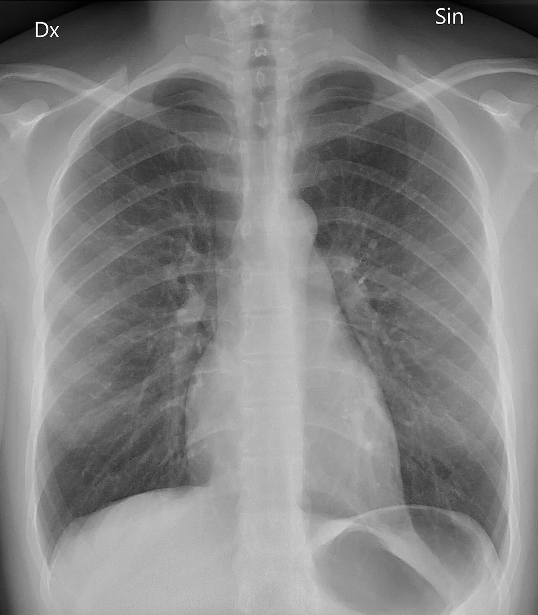
KeeperPA chest: a keeper
Look for: Apices and both costophrenic angles are on the film. The scapulae are outside the lungs, the patient is straight, and the lungs are well inflated.
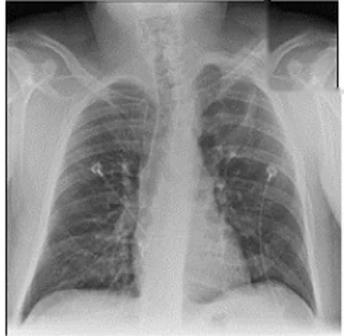
RepeatRotated
Look for: The medial clavicle ends are not the same distance from the spinous process, and the heart and trachea look shifted.
Fix: Square both shoulders against the IR, keep weight even on both feet, and check the shoulders again right before you expose.
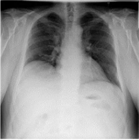
RepeatPoor inspiration
Look for: The diaphragm is high, the lungs look small and crowded, and fewer than 10 posterior ribs show above it.
Fix: Coach the breathing before you go to the control panel. Expose at the end of the second deep breath.
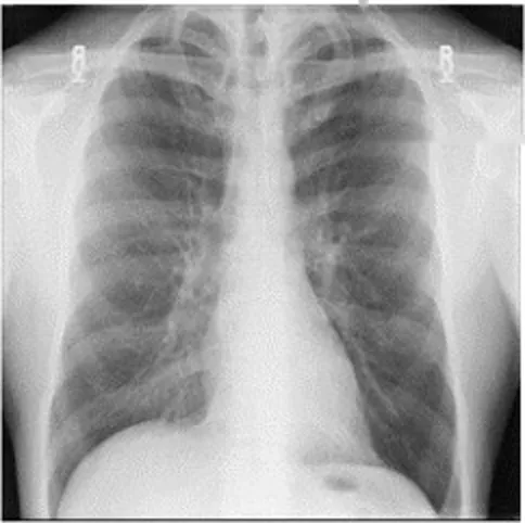
RepeatClipped apices and CP angles
Look for: The tops of the lungs are cut off, and so are both costophrenic angles, where small effusions show first.
Fix: Top of the IR about 1.5 to 2 inches above the shoulders, CR at T7, and open the field enough to include both bases. On a tall patient, turn the IR or use the larger size.
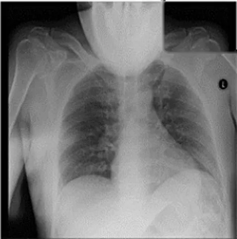
RepeatChin over the apices
Look for: A large rounded soft-tissue shadow covers the apices and the upper airway. It is the patient's chin.
Fix: Raise the chin and rest it on top of the IR before you step out.
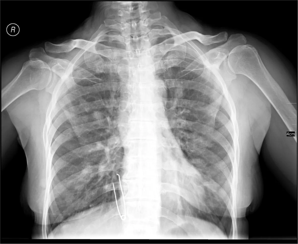
RepeatLeaning back, shoulders not rolled
Look for: The clavicles sit high above the apices and the ribs look flat and horizontal: the patient was leaning back. The scapulae lie over the upper lungs.
Fix: Stand the patient upright against the IR, chin up, and roll the shoulders forward and down.
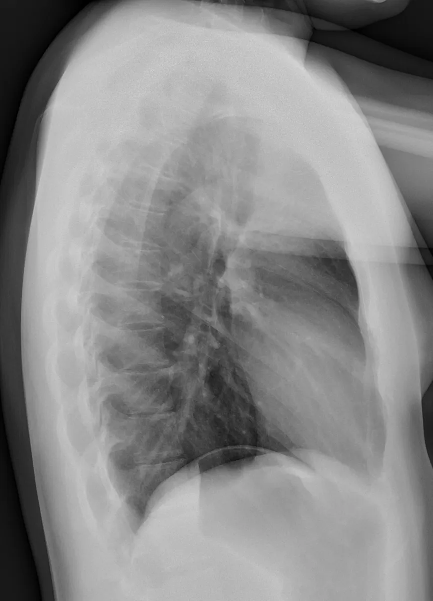
KeeperLeft lateral chest: a keeper
Look for: The posterior ribs line up, the sternum is in profile, and the arms are raised so the upper lungs are clear. The whole lung is included front to back and top to bottom.
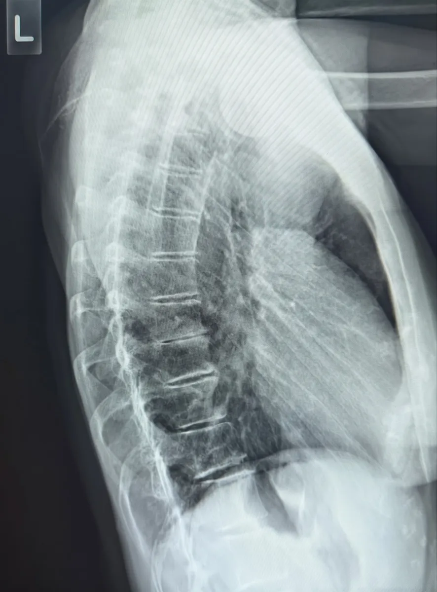
RepeatRotated lateral
Look for: Two separate sets of posterior ribs run down the back of the chest, well over half an inch apart. The patient was turned.
Fix: Make the shoulders and hips perpendicular to the IR. Check from above that the shoulders are stacked.
Clinical Example
Your PA shows 8 posterior ribs and the right clavicle end farther from the spine than the left. Before repeating, you explain the two-breath routine, square the shoulders, and move the patient's weight onto both feet. The repeat shows 10 ribs and clavicle ends that sit evenly.
Common Mistakes
- Checking only the heart and lungs and missing a clipped costophrenic angle.
- Exposing on the first breath.
- Counting anterior ribs instead of posterior ribs.
- Calling up to half an inch of posterior rib separation on a lateral rotation; that much is normal beam divergence.
Why It Matters
Image critique is tested on the registry, and the chest is the film you will critique most at work. Catching a miss before the patient leaves saves a callback and a second trip to the department.
Key Relationships
- Clavicle ends uneven from the spine → rotated
- Fewer than 10 posterior ribs → poor inspiration
- Clavicles high, ribs flat → leaning back (lordotic)
- Scapulae in the lungs → shoulders not rolled forward
- Lateral: posterior ribs more than 1 cm apart → rotated