
Procedures · Head, spine and pelvis2 min read5-question check
Cervical spine
Open mouth for the dens, 72 inches for the lateral, and the oblique angle decides which foramina open.
Patient and part
For the AP axial the patient is supine or erect, not rotated, with the chin raised slightly so the mandible clears the midcervical spine. For the AP open mouth projection of C1 and C2, the mouth is opened wide and the head is adjusted until a line from the lower margin of the upper incisors to the tips of the mastoid processes is perpendicular to the IR. The lateral is taken erect, left or right side against the IR, with both shoulders depressed and the chin slightly raised. For obliques, rotate the body and head 45 degrees.
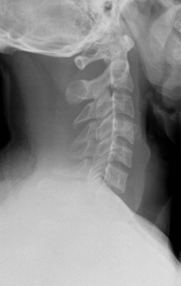
Lateral cervical spine
Central ray
The AP axial uses a 15 to 20 degree cephalad angle centered to C4, at the lower margin of the thyroid cartilage, at 40 inches. The open mouth projection uses a perpendicular CR through the center of the open mouth. The lateral is a horizontal beam to C4 at 72 inches, or 180 cm, because the shoulders create a large OID that the long SID offsets. Obliques also use 72 inches with a 15 to 20 degree angle to C4: caudad for anterior obliques, cephalad for posterior obliques.
What a good image shows
A good AP axial shows open disk spaces from C3 to T2 and midline spinous processes. The open mouth view shows the dens, the body of C2 and the lateral masses of C1 between the teeth and skull base. The lateral shows all seven vertebrae, including C7, and the zygapophyseal joints. Obliques show open, round intervertebral foramina. An anterior oblique shows the foramina closest to the IR, so an RAO shows the right side. A posterior oblique shows the side farthest from the IR, so an RPO shows the left.
Fixing it
On the open mouth view, upper incisors over the dens mean the chin is too low; raise it. The skull base over the dens means the chin is too high; lower it. On the lateral, if C7 is hidden by the shoulders, add a cervicothoracic or swimmer's lateral: the arm nearest the IR is raised overhead and the other shoulder is depressed. Narrowed foramina on an oblique mean the rotation or CR angle was off.
Clinical Example
Your lateral C-spine ends at C6 even though the patient dropped both shoulders. You raise the arm closest to the IR and take a swimmer's lateral centered to T1. C7 and T1 now show between the shoulders.
Common Mistakes
- Using 40 inches for the lateral, which magnifies the spine and blurs it.
- Angling cephalad for an anterior oblique; anterior obliques are angled caudad.
- Mixing up which foramina an oblique shows; posterior obliques show the side up.
- Accepting a lateral that stops above C7.
Why It Matters
Missed C7 injuries and repeats both harm patients. The registry tests the open mouth corrections, the oblique angle direction and which side's foramina each oblique shows.
Key Relationships
- AP axial: 15 to 20 degrees cephalad to C4
- Open mouth: incisors to mastoid tips line perpendicular
- Lateral and obliques: 72 inch SID
- Anterior oblique: caudad angle, foramina nearest the IR
- Posterior oblique: cephalad angle, foramina farthest from the IR