
Image Production · Scatter, grids and filtration2 min read5-question check
Beam Restriction
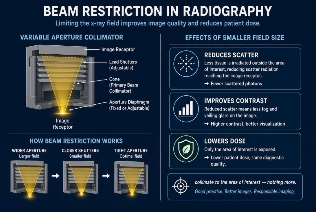
Collimate to the anatomy: less tissue irradiated means less scatter, better contrast, and lower patient dose.
The rule
Beam restriction limits the x-ray field to the anatomy of interest. A smaller field irradiates less tissue, so the patient receives less dose and the body produces less scatter. Less scatter reaching the detector means better image contrast. Few steps improve both dose and image quality at once, which is why collimation is expected on every exam.
Beam Restriction Devices
The devices
The simplest device is the aperture diaphragm, a flat lead sheet with a fixed opening. Cones and cylinders are extensions of an aperture diaphragm that give a fixed circular field. The variable-aperture collimator is the most common device. It has upper shutters near the tube window that reduce off-focus radiation and lower shutters that set the field edges, plus a light and mirror so you can see where the beam will fall. Positive beam limitation, when present, automatically limits the field to the detector size.
The numbers
The light field and the x-ray field must line up within 2% of the SID. Worked example: at a 40-inch SID, 2% is 0.8 inch, about 2 cm. At 72 inches, 2% is about 1.4 inches. If the light field looks centered but the anatomy is clipped on the image, a collimator check by physics or service is needed. Large reductions in field size also reduce scatter reaching the detector, so a tightly collimated large part may need a small mAs increase when using manual technique.
On the job
Collimate before you expose, not afterward. Electronic masking or cropping at the workstation changes only what is displayed; the patient still received the full field dose, and cropping can remove anatomy from the record. Show the collimation borders on the image. Tight collimation also helps the computer read the image correctly, because unattenuated beam outside the part can disrupt histogram analysis.
Clinical Example
For a PA hand, a student opens the field to the full detector. The instructor narrows it to the hand and wrist with a small margin on all sides. The new image has better contrast, a reliable exposure indicator, and a smaller irradiated area.
Common Mistakes
- Opening the field to the full detector size for every exam.
- Using electronic masking to make an uncollimated image look collimated.
- Assuming the light field is always accurate without periodic checks.
- Collimating so tightly that required anatomy is cut off, causing a repeat.
Why It Matters
Collimation is one of the most effective dose and contrast controls a radiographer has. The registry tests devices, the effect on scatter and contrast, and the 2% alignment standard.
Key Relationships
- Smaller field → less scatter → higher contrast
- Smaller field → lower patient dose
- Light and x-ray fields must agree within 2% of SID
- Masking after exposure does not reduce dose