
Image Production · Equipment, AEC and QC2 min read5-question check
Automatic Brightness Control
In fluoroscopy, ABC raises or lowers kVp and mA to hold image brightness steady as anatomy changes.
The rule
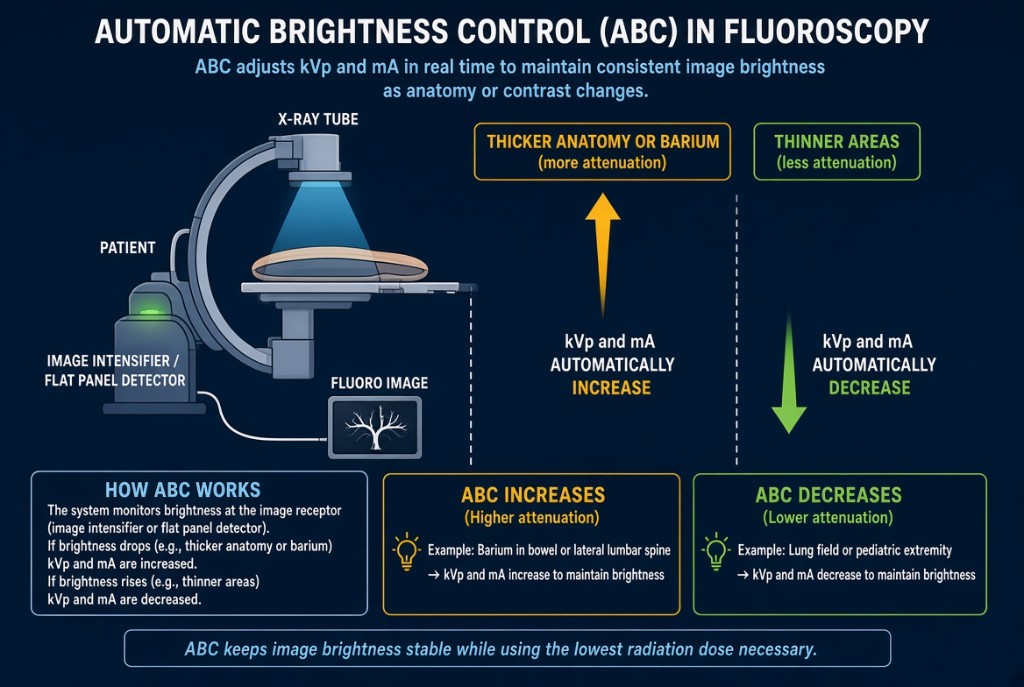
Automatic brightness control, or ABC, is the fluoroscopy feature that keeps the live image at a steady brightness. As the radiologist moves from thin anatomy to thick anatomy, or as barium or iodine enters the field, ABC changes the exposure factors so the image does not go dark or wash out. It is sometimes called automatic dose rate control.
Automatic Brightness Control
How it works
A sensor samples the signal from the image receptor. On an image intensifier, a light sensor reads the output phosphor brightness. On a flat-panel system, the detector signal itself is measured. If the signal drops because the part is thicker or denser, ABC raises kVp, mA, or both. If the signal rises, it lowers them. Different units favor kVp or mA changes depending on the selected program.
The dose
Brightness is held constant by changing the dose. Thick anatomy, a large patient, or a field full of dense contrast drives ABC up, and patient entrance dose rises with it. Federal rules cap the air kerma rate for fluoroscopy units with ABC at 88 mGy per minute in normal mode, and 176 mGy per minute when high-level control is activated. Magnification mode also tends to raise dose, because ABC compensates for the smaller area being imaged.
On the job
Keep the anatomy of interest centered in the field. Metal, lead, or dense contrast near the sensing area can push ABC to high exposure levels. Use the lowest dose mode that gives an adequate image, pulsed fluoroscopy at a low pulse rate when available, last-image hold instead of live fluoro for review, and tight collimation. Watch the cumulative timer and the dose display throughout the case.
Clinical Example
During a barium enema, the radiologist pans over a large patient's pelvis and the dose rate display climbs. The technologist suggests switching from magnification mode back to normal mode and collimating to the rectum. The image stays diagnostic and the dose rate drops.
Common Mistakes
- Assuming a bright, steady image means the dose is low; ABC hides increases in dose.
- Leaving the unit in magnification or high-level mode after it is no longer needed.
- Letting metal or a lead apron drift into the sensing area.
- Thinking ABC is used in standard radiography; that role belongs to AEC.
Why It Matters
ABC protects image quality but can quietly raise patient skin dose during long procedures. The registry tests what ABC adjusts, why dose rises with thick parts, and dose-saving fluoro habits.
Key Relationships
- Thicker or denser part → ABC raises kVp and/or mA
- Constant brightness ≠ constant dose
- Magnification mode → higher patient dose rate
- Normal-mode limit 88 mGy/min; high-level control 176 mGy/min