
Image Production · Geometry and detail2 min read5-question check
Anode heel effect
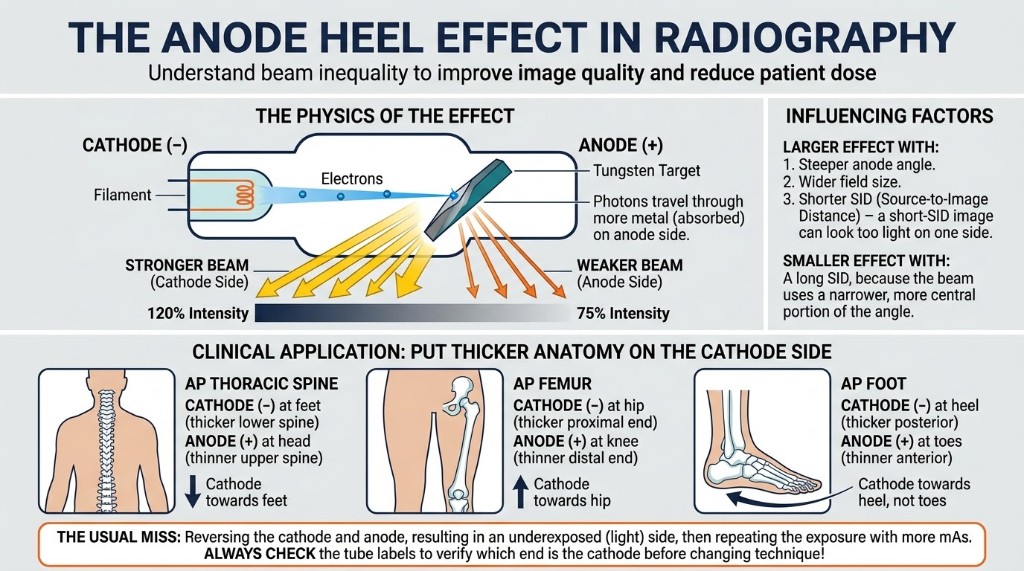
Beam intensity is greater on the cathode side, so put the thicker end of the anatomy under the cathode.
The rule
The x-ray beam is not equally intense across the field. It is stronger on the cathode side and weaker on the anode side. This is the anode heel effect. You use it by placing the thicker or denser end of the anatomy under the cathode, so the stronger part of the beam goes where more penetration is needed and receptor exposure evens out.
Why it happens
The target on the anode is set at an angle, and x-rays are produced slightly below its surface. Photons leaving toward the anode side must pass through more target material, the heel, and some are absorbed. Photons heading toward the cathode side travel through less metal, so fewer are absorbed. The same geometry makes the effective focal spot smaller on the anode side, so spatial resolution is slightly better there.
What makes it worse
The difference is greatest with a smaller target angle, a larger field size, and a shorter SID. A 14 by 17 inch field at 40 inches shows the heel effect clearly. At 72 inches the receptor intercepts only the central part of the beam, where intensity is more even, so a chest shows less of it. Small fields such as a hand show almost none. Long body parts that change thickness from end to end benefit most.
On the job
On an AP thoracic spine, the lower vertebrae sit under a thicker chest, so the cathode goes toward the feet. On an AP femur, the cathode goes toward the hip. On an AP humerus, it goes toward the shoulder. On a forearm, the elbow end goes under the cathode. Check the markings on the tube housing before you position. Digital processing hides some of the unevenness, but noise still increases where fewer photons arrive.
Clinical Example
You are doing an AP femur on a 14 by 17 inch receptor at 40 inches. The stretcher is parked with the patient's hip toward the anode end of the tube. You turn the stretcher so the thick hip lies under the cathode and the thinner knee lies under the anode.
Common Mistakes
- Reversing the tube so the thin end sits under the cathode, which exaggerates the difference.
- Expecting a visible heel effect on small fields such as a hand, where it is negligible.
- Raising mAs to fix one underexposed end, which overexposes the other end instead of evening it out.
- Thinking a longer SID makes the heel effect worse, when it makes it smaller.
Why It Matters
Using the heel effect evens receptor exposure across long body parts with no added dose. The registry asks which end gets the cathode for a given exam and which factors strengthen the effect.
Key Relationships
- Cathode side → greater beam intensity
- Thicker anatomy → under the cathode
- Smaller target angle → stronger heel effect
- Larger field or shorter SID → stronger heel effect
- Anode side → smaller effective focal spot