
Image Production · Equipment, AEC and QC2 min read5-question check
AEC chamber selection
AEC measures only the tissue over the chambers you select, so the anatomy of interest must cover them.
The rule
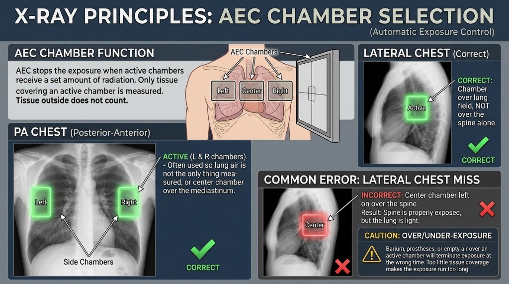
Automatic exposure control ends the exposure once a set amount of radiation has reached the selected detectors, usually ionization chambers placed between the patient and the image receptor. Most systems have three: a center chamber and two outer chambers. The radiographer chooses which ones are active. The AEC only knows about the tissue lying over the active chambers, so the anatomy of interest must completely cover them.
How it works
With AEC you still set kVp, mA, the backup time, and the chamber selection. The system decides the exposure time. Thick or dense tissue over a chamber lets less radiation through, so the exposure runs longer. Thin tissue or open air lets radiation through quickly, so the exposure ends early. A backup timer stops an exposure that runs too long, protecting the patient and the tube.
Wrong chamber
If a chamber sits under air, a thin part, or the edge of the body, it fills quickly and ends the exposure too soon. The anatomy you care about is underexposed and noisy. If a chamber sits under something dense, such as a metal prosthesis, barium in the bowel, or dense tissue that is not the area of interest, the exposure runs long. The anatomy of interest is overexposed and the patient receives more dose than needed. Centering errors cause the same problems, because the right chamber ends up under the wrong anatomy.
On the job
Standard choices follow the anatomy. A PA chest uses the two outer chambers over the lungs. A lateral chest uses the center chamber. An AP lumbar spine uses the center chamber over the vertebrae. Do not use AEC when the part cannot cover the chamber, such as a small extremity or a part near the edge of the field. Accurate centering and tight collimation keep the measurement honest.
Clinical Example
A PA chest is taken with only the center chamber active. The chamber reads through the dense mediastinum and spine, so the exposure runs long, the lungs are overexposed, and the exposure indicator is high. Selecting the two outer chambers over the lungs fixes it.
Common Mistakes
- Leaving the center chamber on for a PA chest, which reads the mediastinum and overexposes the lungs.
- Letting a chamber sit under air beside an off-center patient, which ends the exposure early.
- Thinking a prosthesis over the chamber shortens the exposure, when it lengthens it.
- Using AEC for a part too small to cover the chamber.
Why It Matters
Correct chamber selection is what makes AEC consistent, and the wrong choice causes repeats and extra dose. The registry gives a scenario and asks which chamber to use or why the exposure came out high or low.
Key Relationships
- Dense material over chamber → longer exposure → overexposure
- Air or thin tissue over chamber → early termination → underexposure
- PA chest → two outer chambers; lateral chest → center chamber
- AEC controls exposure time; you still set kVp